Join us for a BEP/MEP project!
We regularly have projects for (applied) physics and nanobiology students, but also ran projects with students in biomedical engineering, life science and technology, and molecular science and technology. Below are some example projects, please contact Jacob for up-to-date possibilities matched to our current research efforts.
We regularly have projects for (applied) physics and nanobiology students, but also ran projects with students in biomedical engineering, life science and technology, and molecular science and technology. Below are some example projects, please contact Jacob for up-to-date possibilities matched to our current research efforts.
"Google Maps" for the brain
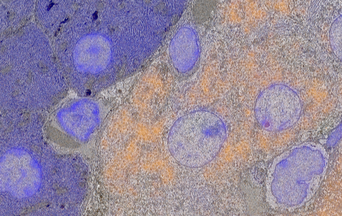
With electron microscopy a 'google maps' can be build, e.g., to map all connections in a brain. However with resolution of one to a few nanometers, throughput is very low and in the grayscale images proteins and other biological molecules are not visible. We are developing new techniques based on parallel scanning with multiple beams, integration of light and electron microscopy, and development of dedicated probes and labels to allow fast, high-resolution imaging over many length scales. In this way we can zoom in and out on tissue from millimetres to molecules. Student projects can involve experimental work, wet bench work, programming, data and image analysis, and machine learning.
With electron microscopy a 'google maps' can be build, e.g., to map all connections in a brain. However with resolution of one to a few nanometers, throughput is very low and in the grayscale images proteins and other biological molecules are not visible. We are developing new techniques based on parallel scanning with multiple beams, integration of light and electron microscopy, and development of dedicated probes and labels to allow fast, high-resolution imaging over many length scales. In this way we can zoom in and out on tissue from millimetres to molecules. Student projects can involve experimental work, wet bench work, programming, data and image analysis, and machine learning.
Timing escape times of photons
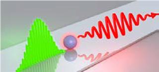
Our group has built a unique microscope that can determine the time it takes for photons to escape from a material. The photons are generated when an electron beam hits a (nano)particle. The escape time bears unique knowledge about the particle and its local environment. The student is asked to do experiments to measure photon escape times from nanoparticles in different environments.
Our group has built a unique microscope that can determine the time it takes for photons to escape from a material. The photons are generated when an electron beam hits a (nano)particle. The escape time bears unique knowledge about the particle and its local environment. The student is asked to do experiments to measure photon escape times from nanoparticles in different environments.
Watching slow electrons at work
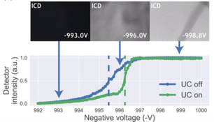
Slow electrons (energy of 0-20eV) are generated under high energy irradiation (e.g., e-beam, EUV) and diffuse and react in the irradiated material. Understanding these processes is very important for high-resolution microscopy and lithography. We have invented a technique to generate these slow electrons and watch their diffusion and reactions in an exposed material. Students projects can involve work on experimental setup, electron microscopy, measurements and data analysis, numerical simulations
Slow electrons (energy of 0-20eV) are generated under high energy irradiation (e.g., e-beam, EUV) and diffuse and react in the irradiated material. Understanding these processes is very important for high-resolution microscopy and lithography. We have invented a technique to generate these slow electrons and watch their diffusion and reactions in an exposed material. Students projects can involve work on experimental setup, electron microscopy, measurements and data analysis, numerical simulations
Printing molecules at the nanoscale
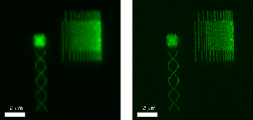
Superresolution techniques achieve light microscopy with a resolution far below the diffration limit. Testing, calibrating, and evaluating the performance of these microscopes is highly challenging because a suitable test sample needs molecules at precisely defined nanoscale postions. Together with colleagues at Erasmus MC, we have optimized a technique using electron beam exposure followed by antibody-functionalization, to print fluorescent molecules at sub-100 nm length scales. Student projects involve electron microscopy, wet-bench work, (superresolution) fluorescence microscopy, and data analysis.
Superresolution techniques achieve light microscopy with a resolution far below the diffration limit. Testing, calibrating, and evaluating the performance of these microscopes is highly challenging because a suitable test sample needs molecules at precisely defined nanoscale postions. Together with colleagues at Erasmus MC, we have optimized a technique using electron beam exposure followed by antibody-functionalization, to print fluorescent molecules at sub-100 nm length scales. Student projects involve electron microscopy, wet-bench work, (superresolution) fluorescence microscopy, and data analysis.
Superresolution with an electron beam
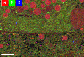
Superresolution fluorescence has revolutionized light microscopy by imaging with a resolution an order of magnitude or more below the diffraction limit. Electron microscopy intrinsically has a resolution of about a nanometer, but may be hard to combine with standard superresolution fluorescence techniques. We are inventing new techniques using the electron beam to do very high resolution fluorescence light microscopy and we collaborate with microscopists in medical centers to apply these techniques on their biomedical samples. Student projects can involve construction and design of experimental set-up, experimental work, data analysis, and numerical simulations.
Superresolution fluorescence has revolutionized light microscopy by imaging with a resolution an order of magnitude or more below the diffraction limit. Electron microscopy intrinsically has a resolution of about a nanometer, but may be hard to combine with standard superresolution fluorescence techniques. We are inventing new techniques using the electron beam to do very high resolution fluorescence light microscopy and we collaborate with microscopists in medical centers to apply these techniques on their biomedical samples. Student projects can involve construction and design of experimental set-up, experimental work, data analysis, and numerical simulations.
Finding a protein in a cellular haystack
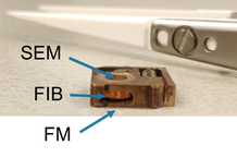
With FIB-SEM proteins of interest can be carved out of a cell for high-resolution cryo-TEM. However, finding the protein of interest inside a cell is like finding a needle in a haystack. We want to achieve 3D fluorescence localization inside a (cryo) FIB-SEM to pin-point the protein. The focused ion beam is then used to mill out a thin lamella containing the protein. Student projects can involve instrumentation, experimental work, optics, and numerical simulations.
With FIB-SEM proteins of interest can be carved out of a cell for high-resolution cryo-TEM. However, finding the protein of interest inside a cell is like finding a needle in a haystack. We want to achieve 3D fluorescence localization inside a (cryo) FIB-SEM to pin-point the protein. The focused ion beam is then used to mill out a thin lamella containing the protein. Student projects can involve instrumentation, experimental work, optics, and numerical simulations.